Tools & Techniques
Alongside biological projects, my group develops and applies methods that make membrane-protein structural biology more tractable and interpretable, through a mix of in-house work and external collaborations. This includes engineered binders for crystallisation and cryo-EM, high-throughput expression and purification pipelines, biosensors for functional readouts, and mass-spectrometry approaches to protein dynamics such as HDX-MS (hydrogen-deuterium exchange) and carbene footprinting, which resolve conformational changes that static structures cannot capture.
Nanobodies and binders
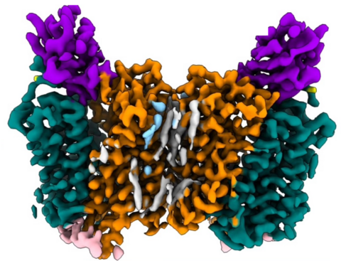
- Protein-binder toolbox for solute carriers (J Mol Biol, 2024) — 525 binders against 22 SLCs, validated with a cell-based pipeline (immunofluorescence, immunoprecipitation) and applied to structural studies on KCC3b (SLC12A6); cryo-EM maps EMD-50364, 50405, 50406, 50407.
- Gluebodies: transferable nanobody mutations for reliable crystallisation (ACS Cent Sci, 2025)
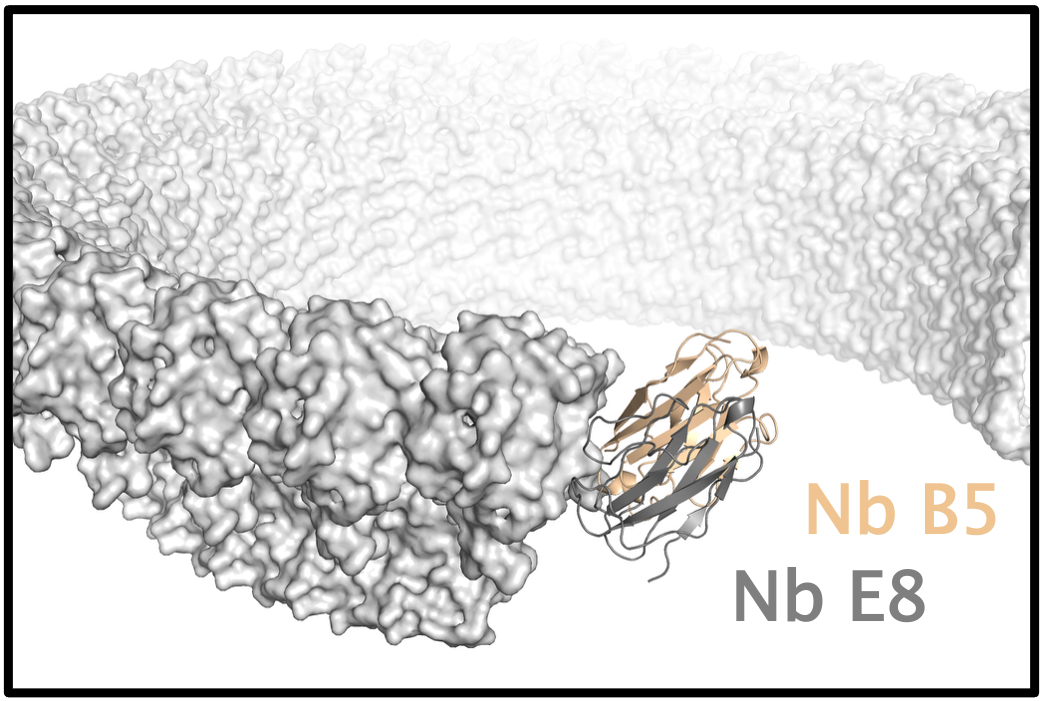
- Anti-gasdermin D nanobodies (Nb B5, Nb E8) that bind gasdermin D and prevent pore formation
- Anti-BRIL nanobody — a nanobody against the BRIL (apocytochrome b562RIL) fusion domain, as an affinity reagent for purification of BRIL-tagged constructs
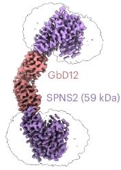
- Di-Gembodies: covalent nanobody dimers for cryo-EM of small proteins (Nat Chem Biol, 2025)
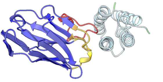
- Crystal structure of the Tspan15 LEL domain and its ADAM10 binding site (Structure, 2022) — enabled by Fab 1C12
Mass spectrometry for protein dynamics
We use HDX-MS and carbene footprinting to map ligand binding sites and conformational change, complementing static structural data from cryo-EM and X-ray crystallography.
- Native MS and HDX-MS of the MC2R–MRAP complex and antagonist-driven conformational change (bioRxiv, 2026)
- Multi-region carbene footprinting of the β1-adrenergic receptor (Chem Sci, 2025)
- Carbene footprinting of Gasdermin D and Caspase-1 binding (JACS Au, 2023)
High-throughput expression and biochemistry
- High-throughput expression and purification of human SLCs (JoVE, 2023)
- Expression screening of integral membrane proteins using BacMam (Methods Mol Biol, 2020)
Target Enabling Packages (SGC)

Public Target Enabling Packages produced through the SGC Membrane Protein Structure & Function group, bundling reagents, assays and structural information for open use by the community.
- SLC12A4 (KCC1) and SLC12A6 (KCC3) Target Enabling Package, v1 (2024) (PDF, thesgc.org)
Functional readouts and electrophysiology
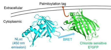
- BRET-based chloride biosensor for high-throughput KCC2 screening (SLAS Discov, 2025)
- Cation transport measured by atomic absorption in Xenopus oocytes (JoVE, 2013)
- Voltage-clamp fluorometry of the gastric H,K-ATPase (PLoS ONE, 2012)
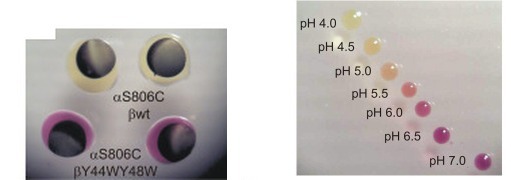
- Acidification assay for proton transport by the gastric H,K-ATPase in Xenopus oocytes (JBC, 2009)